By Dr. Petrus Raulino
A study published in the journal Frontiers in Psychiatry brings some more possible pieces of the great puzzle that is doing research on the biological bases of depression.
The study showed differences between the cellular composition of the brain in depressed adults who died by suicide compared to individuals with no psychiatric history who died suddenly by other means.
Analysis of the study
Post-mortem brain samples from males who died by suicide in the presence of a severe depressive episode and brain samples from males who died suddenly without any inflammatory, psychiatric or neurological illness were analyzed.
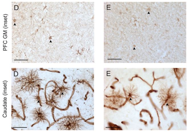
Three brain regions were analyzed in all the study subjects: the dorsomedial prefrontal cortex, the dorsal caudate nucleus and the mid-dorsal thalamus. Astrocytes were analyzed in these regions.
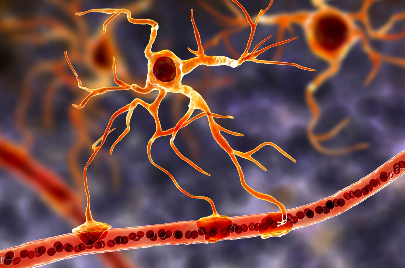
Astrocytes are often star-shaped glial cells that make up approximately half of the cells in the human brain.
Astrocytes form the structure of the brain and have important functions such as supporting and nourishing neurons.
Astrocytes are heterogeneous cells, with different subtypes in terms of morphology, development, metabolism and physiology.
Astrocytes in the brain were analyzed by staining specific proteins found in their structure (glial fibrillary acidic protein and vimentin).
Using a microscope, the number of astrocytes was counted in cross-sections.
The vascular densities of the brain regions included in the study were also assessed, as a previous study found a close inverse association between regional astrocyte density and vascular density.
Main results of the survey
The research showed a reduction in the number of astrocytes in the brain areas of depressed adults.
The vascular density and morphometric characteristics of the astrocytes were similar between the groups, except in the prefrontal white matter, where there was increased vascularization and the astrocytes exhibited fewer primary processes (extensions).
Although the structure of astrocytes in depressed individuals was similar to that of individuals without a psychiatric history, the number of astrocytes was reduced in depression.
Some limitations of the research were important to highlight:
- small sample;
- no women in the sample;
- lack of information on the number and severity of previous depressive episodes;
- evolution time;
- duration of the current episode;
- treatments, etc.
Conclusion
In any case, the study is thought-provoking enough to stimulate further research.
Could depression be linked to the cellular composition of the brain? If so, is it static or dynamic?
Perhaps the most promising news in the future will be the knowledge that, unlike neurons, the adult human brain continually produces new astrocytes.
References
O'Leary, L. A., Belliveau, C., Davoli, M. A., Ma, J. C., Tanti, A., Turecki, G., & Mechawar, N. (2021). Widespread decrease of cerebral vimentin-immunoreactive astrocytes in depressed suicides. Frontiers in Psychiatry, 12, 75.
O'Leary, L. A., Davoli, M. A., Belliveau, C., Tanti, A., Ma, J. C., Farmer, W. T., ... & Mechawar, N. (2020). Characterization of vimentin-immunoreactive astrocytes in the human brain. Frontiers in Neuroanatomy, 14.