By Dr. Petrus Raulino
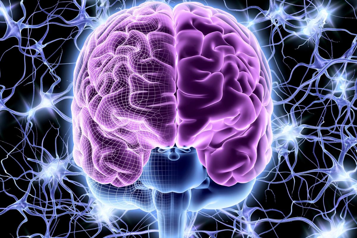
What is Synaptic Pruning?
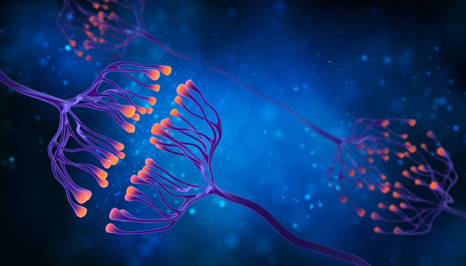
Synaptic pruning is the natural process of eliminating synapses that were produced "in excess" in the first years of life. It usually occurs more rapidly in childhood and adolescence, with a gradual decrease until early adulthood.
The number of synapses in an adult is about half that of a child.
Synaptic pruning can be understood as fine-tuning the nervous system by deleting extra synapses.
The process of synaptic pruning overlaps in time with the age of onset of many cases of schizophrenia, which is late adolescence to early adulthood.
Synaptic Pruning in Schizophrenia
In schizophreniaIt seems that the normal process of synaptic pruning is disrupted, causing widespread damage to neuronal communication.
It is therefore hypothesized that the excessive loss of functional synapses, rather than the normal refinement of circuits, leads to the development of schizophrenia.
There is a lot of evidence of synaptic dysfunction in patients with schizophrenia.
Post-mortem evidence has shown a significant reduction in synaptophysin in patients with schizophrenia compared to the control group. Synaptophysin is a glycoprotein that serves as a marker of synaptic density.
In patients with schizophrenia, synaptic density tends to be lower in the frontal cortex and anterior cingulate cortex.
But what is the hypothesis to explain the mechanism of loss of synaptic density in schizophrenia?
Evidence suggests that altered microglial cell activity can lead to dysregulated synaptic pruning in cortical regions, causing cognitive problems and symptoms associated with schizophrenia.
If this is the case, pharmacological interventions that target the microglial elimination of synapses deserve further research with the aim of seeking therapeutic options to protect the brains of individuals at risk of schizophrenia.
References
Onwordi, E. C., Halff, E. F., Whitehurst, T., Mansur, A., Cotel, M. C., Wells, L., ... & Howes, O. D. (2020). Synaptic density marker SV2A is reduced in schizophrenia patients and unaffected by antipsychotics in rats. Nature communications, 11(1), 1-11.
McCutcheon, R. A., Marques, T. R., & Howes, O. D. (2020). Schizophrenia-an overview. JAMA psychiatry, 77(2), 201-210.
Osimo, E. F., Beck, K., Marques, T. R., & Howes, O. D. (2019). Synaptic loss in schizophrenia: a meta-analysis and systematic review of synaptic protein and mRNA measures. Molecular psychiatry, 24(4), 549-561.
Sellgren, C. M., Gracias, J., Watmuff, B., Biag, J. D., Thanos, J. M., Whittredge, P. B., ... & Perlis, R. H. (2019). Increased synapse elimination by microglia in schizophrenia patients-derived models of synaptic pruning. Nature neuroscience, 22(3), 374-385.
Howes, O. D., & McCutcheon, R. (2017). Inflammation and the neural diathesis-stress hypothesis of schizophrenia: a reconceptualization. Translational psychiatry, 7(2), e1024-e1024.
Finnema, S. J., Nabulsi, N. B., Eid, T., Detyniecki, K., Lin, S. F., Chen, M. K., ... & Carson, R. E. (2016). Imaging synaptic density in the living human brain. Science translational medicine, 8(348), 348ra96-348ra96.
Sekar, A., Bialas, A. R., De Rivera, H., Davis, A., Hammond, T. R., Kamitaki, N., ... & McCarroll, S. A. (2016). Schizophrenia risk from complex variation of complement component 4. Nature, 530(7589), 177-183.
Selemon, L. D., & Zecevic, N. (2015). Schizophrenia: a tale of two critical periods for prefrontal cortical development. Translational psychiatry, 5(8), e623-e623.
Stephan, A. H., Barres, B. A., & Stevens, B. (2012). The complement system: an unexpected role in synaptic pruning during development and disease. Annual review of neuroscience, 35, 369-389.
Paus, T., Keshavan, M., & Giedd, J. N. (2008). Why do many psychiatric disorders emerge during adolescence? Nature reviews neuroscience, 9(12), 947-957.